Oral Surgery
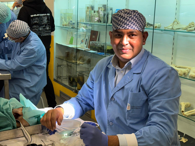
Dr Najeeb Hussain
Our Principal, Dr Najeeb Hussain, is a member of the British Association of Oral and Maxillofacial Surgeons. Dr Hussain has been practicing for more than 25 years with special interest in Oral Surgery, Dental Implants and Conscious Sedation. Visit our Team page to find out more about his expertise.
- At Morden Dentist we accept private referrals for the following oral surgery procedures: wisdom teeth extraction, frenectomies, tooth exposures and apicoectomies.
- We also accept NHS patients who are referred to us for oral surgery, under a separate set of NHS rules. Morden Dentist is the referral practice for intermediate minor oral surgery (IMOS) for the London Boroughs of Sutton & Merton. Please visit our NHS IMOS page for more information.
Oral Surgery Procedures
Wisdom Teeth
Wisdom teeth are the third-last permanent molars. Most people have four wisdom teeth, two in the upper jaw and two in the lower jaw. These teeth are commonly called wisdom teeth because they usually erupt between the ages of 16 to 21, known as the ‘age of wisdom’. A wisdom tooth is impacted when it is obstructed from erupting fully into the mouth by the tooth in front of it or the surrounding bone or gums.
Problems caused by impacted wisdom teeth
Improperly erupted wisdom teeth are breeding grounds for bacteria and may cause tooth decay, sometimes even affecting the neighbouring teeth. Infection of the overlying gums can take place as well, resulting in pain and swelling.
More serious problems such as the formation of cysts or tumours around an impacted tooth can occur, leading to destruction of the surrounding jawbone and neighbouring teeth. These conditions may require complex and extensive treatment. As problems can develop silently, without your knowledge, a check-up with your dentist is advisable.
Wisdom teeth consultation
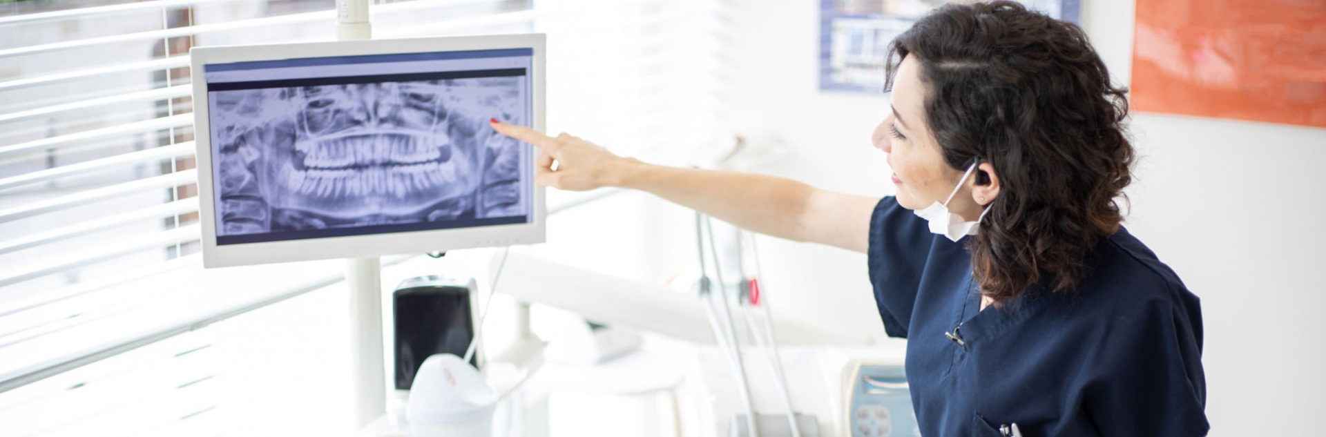
- Your initial visit to Morden Dentist will include an examination of your mouth and X-rays to determine the position of the wisdom teeth, their condition and the status of the adjacent teeth and bone.
- To prevent problems associated with impacted wisdom teeth, it’s advisable to remove them early. The best time to remove them would be during the teenage years, before the roots of the teeth are fully formed and firmly embedded in the jawbone. Healing is also better during this period, with less risk of complications.
- This is a minor surgical procedure that can usually be performed with little discomfort. The procedure can be performed under local anaesthesia (with or without sedation to control anxiety) or general anaesthesia. Dr Najeeb Hussain will advise you on the type most appropriate for your needs.
- The surgery involves uncovering the tooth by lifting the overlying gums aside to expose the tooth and bone. The tooth may need to be sectioned in order to remove it. The gums are then stitched back.
Wisdom Teeth FAQs
Although most people develop and grow 32 permanent, adult teeth (16 in the upper and 16 in the lower jaw), many do not have enough room in their mouth for all of these teeth to completely erupt. Since the wisdom teeth are the last to develop, they will not always have enough room to adequately erupt into the mouth to become fully functional teeth. This lack of room or space can result in a number of harmful effects on your overall dental health. When this occurs they are said to be impacted, indicating their inability to erupt into an alignment which would allow them to function in the chewing process.
If it is recognised that you do not have enough room in your mouth for your third molars to erupt, it is advisable to have them removed as soon as possible. In some patients, this may be as early as 17 or 18 years of age. Early removal allows for faster healing, more predictable final results, and fewer complications compared to older patients.
After surgery, some minor bleeding from the wound can be expected, which can be controlled by biting on a piece of gauze over the operated area for about half an hour. Facial swelling and discolouration of the overlying skin will also develop, increasing for the first 72 hours and subsiding thereafter. You may not be able to open your mouth as wide as usual for a few days.
Painkillers, antibiotics and an antiseptic mouthwash are usually prescribed after the surgery. You will be advised to maintain good oral hygiene and also to keep to a soft diet for a few days following surgery.

Frenectomies
A frenectomy is simply the removal of a frenum in the mouth. A frenum is a muscular attachment between two tissues. There are two frena (the plural form of frenum) in the mouth that can sometimes obstruct normal function and are candidates for frenectomies. These frena are called the lingual frenum, which connect the tongue to the floor of the mouth, and the maxillary labial frenum, which connects the inside of your upper lip to your gums just above your upper two front teeth.
The Frenectomy procedure is a simple surgical procedure that generally takes less than thirty minutes to complete. The procedure is performed using local anaesthesia, sometimes with sedation as well.
Frenectomies FAQs
Frenectomies will usually take a couple of weeks to completely heal. You may have to take over-the-counter non-steroidal anti-inflammatory (NSAID) drugs like Ibuprofen to relieve any pain that you may have.
You should rinse with saltwater or an antiseptic mouthwash such as Chlorhexidine mouthwash in order to keep the surgical area clean. You should brush carefully around the area and floss daily as well.
If you have had Frenectomy using a scalpel, you will have absorbable sutures which should disappear by themselves within a couple of weeks. You should come back to see us after two weeks to remove any sutures that have not resorbed and for Dr Najeeb Hussain to check good healing has occurred.
Like many other surgical procedures, there can be some pain, swelling, bruising, tenderness, gum scarring and bleeding after Frenectomy surgery. If the Frenectomy operation does not solve the problem completely then it may have to be repeated. Occasionally nerve damage may occur after Frenectomy surgery.
This damage may be in the form of a slight tingling sensation around the surgical area to total numbness of the area. Nerve damage in this area may only be temporary, and after a few weeks, sensations in the area may increase. On rare occasions, the nerve damage persists, and feeling around the surgery area is not regained.
Tooth Exposures
When a tooth fails to emerge through the gums, it’s considered to be an impacted tooth. This commonly occurs in the case of canine teeth. It’s important to treat an impacted tooth in order to prevent the improper eruption of nearby teeth, cyst formation, possible infection or other negative changes in the jaw.
In order to determine the correct treatment for you, Dr Najeeb Hussain will examine your teeth and radiographs to determine the position of the impacted tooth as well as the condition of your gums. One of the treatment options is to surgically expose the tooth in order to guide it to the right position in your jaw. In most cases, you will need to have space created in your jaw through orthodontics prior to the surgical treatment.

Tooth Exposures FAQs
Surgically exposing and aligning impacted canines can prevent further damage to your dentition. In conjunction with subsequent orthodontic treatment, it can result in a more aesthetically pleasing appearance.
Depending on the location, there are different methods of accomplishing the surgical exposure of your tooth. If the impacted canine is close to the palate, Dr Najeeb Hussain will have two options depending on the exact position of the tooth.
The first option is to expose the tooth in order to allow it to erupt on its own. Once the surgery is completed, we may place a protective dressing over the surgical site while it heals. This method will allow the canine tooth to emerge until it’s at the level of the adjacent teeth, after which the teeth can be aligned with braces. The second option is to expose the tooth and then attach an orthodontic bracket to it either at the time of surgery or shortly after in order to help guide the tooth to the level of the adjacent teeth.
If the impacted canine is close to the outer, facial, aspect of the upper jaw, depending on the exact position of the tooth and the condition of your gums, there are 3 different treatment options we can offer:
Option 1: in the first option, we’ll expose the tooth and reposition your gums so as to leave some of the crown of the tooth exposed. We’ll then attach an orthodontic bracket to the tooth and use it to guide the tooth to its proper position.
Option 2: the second option is called a closed technique because, after we expose the tooth, we’ll attach a bracket to it and then replace your gums back to their original position. Only the orthodontic wire will be visible through your gums while the tooth is guided to its proper position. Once your orthodontic treatment is completed, minor recontouring of your gums may be necessary.
Option 3: in the third method, we’ll will create a window through the gums to the surface of the tooth. We’ll then attach an orthodontic bracket to the tooth to help guide it to its proper position. Following orthodontic treatment, we’ll place a gum graft at the neck of the tooth in order to replace any missing gum tissue.
If stitches were used during your surgical procedure, you may need to have them removed 1 to 2 weeks post-operatively. Regardless of which treatment you have received, you must avoid chewing on the surgical site for 2 weeks following your surgery. If prescribed, you must rinse with Chlorhexidine twice daily for 2 minutes until the surgical site is comfortable and you can resume good dental hygiene, which is, brushing and flossing daily. At the time of surgery, we’ll provide you with the appropriate pain medication, or will prescribe pain medication for the post operative period.
Apicoectomies
Your teeth are held in place by roots that extend into your jawbone. Front teeth usually have one root, whilst other teeth, such as your premolars and molars, have two or more roots. During root canal treatment, the canals are cleaned, and inflamed or infected tissue is removed.
Root canals are very complex, with many small branches off the main canal and sometimes, even after root canal treatment, infected debris can remain in these branches and possibly prevent healing or cause re-infection later.
If an infection develops or persists after root canal treatment or retreatment, an apicoectomy may be needed. In an apicoectomy, the root tip, or apex, is removed along with the infected tissue. A filling is then placed to seal the end of the root.
An apicoectomy is sometimes called endodontic microsurgery because the procedure is done under an operating microscope. Dr Najeeb Hussain can do an apicoectomy to fix the problem so the tooth doesn’t need to be extracted. An apicoectomy is done only after a tooth has had at least one root canal procedure. An apicoectomy is not the same as a root resection. In a root resection, an entire root is removed, rather than just the tip.
Apicectomies FAQs
We may take x-rays and you may be given an antimicrobial mouth rinse, anti-inflammatory medication and/or antibiotics before the surgery.
The endodontist will cut and lift the gum away from the tooth so the root is easily accessible. The infected tissue will be removed along with the last few millimetres of the root tip. We use a dye that highlights cracks and fractures in the tooth. If the tooth is cracked or fractured, it may have to be extracted, and the apicectomy will not continue.
To complete the apicectomy, 3 to 4 millimetres of the tooth’s canal are cleaned and sealed. The cleaning is usually done under a microscope using ultrasonic instruments. Use of a surgical microscope increases the chances for success because the light and magnification allow the endodontist to see the area better. Your endodontist will take an X-ray of the area before suturing the tissue back in place.
Most apicoectomies take between 30 to 90 minutes, depending on the location of the tooth and the complexity of the root structure. Procedures on front teeth are generally the shortest. Those on lower molars generally take the longest.
You’ll receive instructions from your endodontist about which medications to take and what you can eat or drink. You should ice the area directly outside the cheek, intermittently (for no longer than 10-15 minutes at a time) for about 12 hours after the surgery, and rest during that time.
The area may bruise and swell. It may be more swollen the second day after the procedure than the first day. Any pain can usually be controlled with over-the-counter nonsteroidal anti-inflammatory drugs (NSAIDs), such as ibuprofen (Advil, Motrin and others) or prescription medication.
To allow for healing, you should avoid brushing the area, rinsing vigorously, smoking or eating crunchy or hard foods. Do not lift your lip to examine the area, because this can disrupt blood-clot formation and loosen the sutures. You may have some numbness in the area for days or weeks from the trauma of the surgery. This does not mean that nerves have been damaged. Tell us about any numbness you experience.
Your stitches will be removed 5 to 7 days after the procedure, and all soreness and swelling are usually gone by 14 days after the procedure. Even though an apicectomy is considered surgery, many people say that recovering from an apicectomy is easier than recovering from the original root-canal treatment.
If you’re having any pain or swelling following root-canal treatment, contact us and we’ll take X-rays and do an examination, to see if an apicectomy is advisable.
If you would like to know more about Oral Surgery at Morden Dentist, please call us today or fill in our enquiry form below.